


If you or someone you love is living with unexplained symptoms, the search for answers can feel long, frustrating and uncertain. This is where a skin biopsy can make a real difference.
A skin biopsy might sound daunting, but in reality, it’s a simple and powerful way for doctors to gather vital clues about what’s happening inside the body. Just beneath the surface of the skin lies a network of tiny nerve fibres and cells that can reveal important information about certain conditions, including some rare and complex diseases.
By examining a very small sample of skin under a microscope, specialists can begin to see patterns and changes that aren’t visible through scans or blood tests. For many patients and families, this test can bring them one step closer to understanding the cause of symptoms and finding the right support or treatment.
But what actually happens to that tiny sample once it leaves the clinic? How can it help researchers uncover what might be going wrong inside the body?
In this guest blog, Newcastle PhD student Alice Smail, who’s involved in the LifeArc Centre for Rare Mitochondrial Diseases, takes us inside the lab to show how skin biopsies are used in mitochondrial research, and how techniques like western blotting can help turn small samples into meaningful answers.
What happens to skin biopsies in mitochondrial research?
Skin biopsies are really useful for researchers, as we can perform lots of different experiments on them to investigate mitochondrial disease. One of these experiments is a western blot. But before we learn what a western blot is, let’s cover some background questions...
What is a cell?
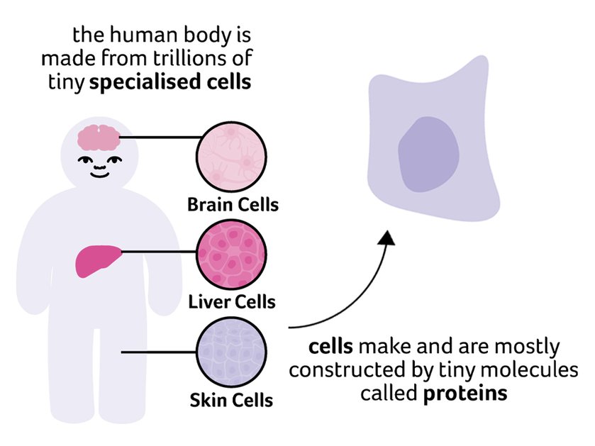
The human body is made up of specialised cells that carry out different functions. Cells are mostly formed from tiny molecules called proteins. Usually ‘protein’ refers to the protein in food – for example, beans contain a lot of protein – but when biologists say protein they’re referring to the tiny individual proteins created within cells. Different cell types contain different levels of these proteins, which makes them uniquely specialised to their role in the body.
What are proteins?
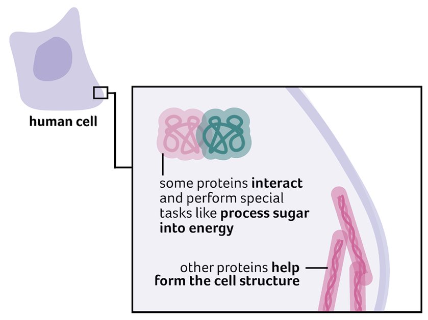
Proteins are chains of chemicals that form different shapes. Some proteins are like long scaffolds that form the cell structure, while others are rounded blobs that help the cell digest other chemicals. A lot of proteins stay inside the cell, but others are secreted and go elsewhere in the body. For example, you’ve probably heard of insulin, which helps cells take up sugar from the blood for energy: insulin is a protein made and secreted by specialised cells in the pancreas.
What is DNA?
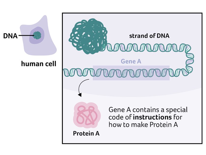

DNA is found in almost every cell, inside a special compartment called the nucleus. A stretch of DNA that contains instructions to make a specific protein is called a gene: the genetic code in a gene tells the cell how to make the protein (there are also some genes that encode other things the cell needs). The human genome is the full list of DNA that contains instructions for making all of the 20,000 proteins that are needed to make a human. It’s a bit like a recipe book containing different ingredients for each protein! Most cells contain two copies of the whole recipe book (the whole genome), but different cells use different recipes (genes).
Are proteins important for mitochondria?
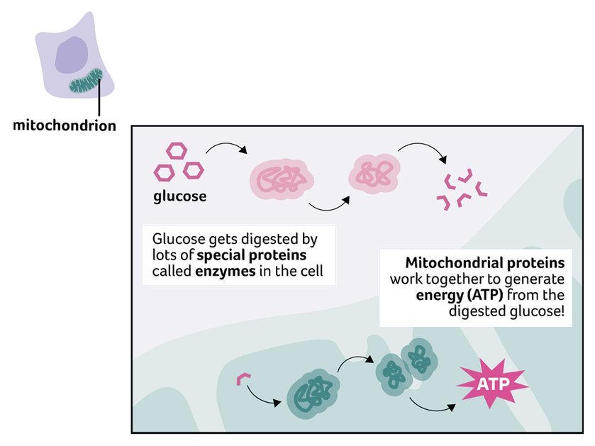
Yes! Mitochondria are found in most cells and they convert sugar into units of energy (called ATP) that the cell can use. This is carried out by different proteins working together. Mitochondria have other important roles, which are also controlled by different groups of proteins working together: in total, over a thousand proteins are needed for mitochondria to work properly.

Why do researchers use western blots?
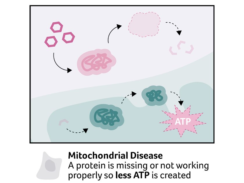
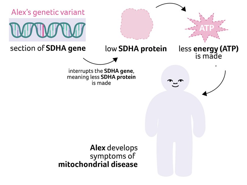
If a mitochondrial protein is missing, the mitochondria might not create enough energy, and the cell won’t function properly, causing the symptoms of mitochondrial disease. Researchers might think that someone has mitochondrial disease because they have a genetic variant that interrupts the instructions (the gene) for a specific protein. To investigate this, researchers can use a western blot to look at protein levels.
Case study
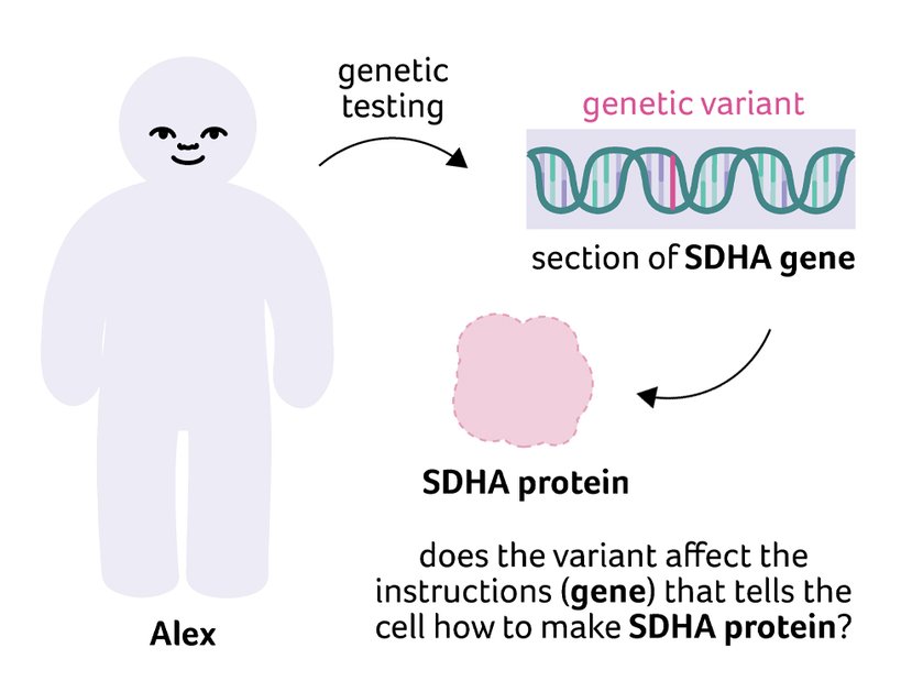
Alex is a 10-year-old boy who has some symptoms of mitochondrial disease. Results from genetic testing show that he carries a genetic variant in both copies of his SDHA gene. The SDHA gene contains the recipe for a mitochondrial protein (also called SDHA) that helps make energy (ATP). However, the doctors have never seen this variant before so they don’t know whether they can provide Alex with a genetic diagnosis yet. They take a skin biopsy from Alex to investigate further.
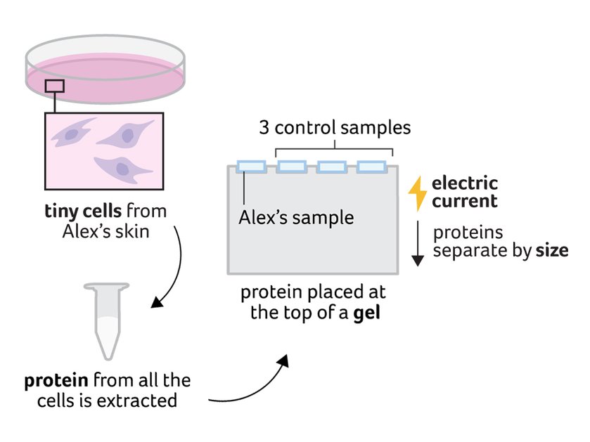
In the lab, a researcher grows cells from Alex’s skin biopsy in a small plastic dish. After the cells have grown for a few days, the researcher extracts all of the protein in the cells using chemicals and heat. Then, they begin the first step of a western blot. A small amount of the protein mix is placed on top of a thin jelly-like gel. The researcher also puts the same amount of protein from control cells (cells from someone without mitochondrial disease) next to Alex’s sample on the gel. An electric current makes the proteins move towards the bottom of the gel. Larger proteins move more slowly, so they stay near the top of the gel, while small proteins move faster and nearly reach the bottom.
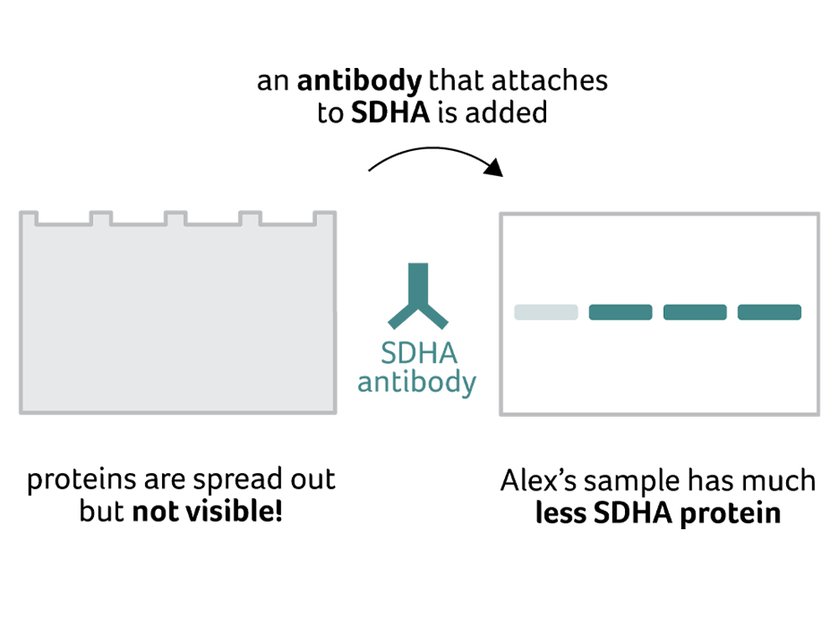
Once this is complete, the researcher has a gel with proteins spread across it according to their size. Next the researcher moves all the proteins onto a special papery membrane. This membrane is soaked in antibodies that cling to SDHA protein; if there’s a lot of SDHA in the sample, lots of antibodies attach. The researcher can view the concentration of attached antibodies using a special camera. In Alex’s sample, there’s not very much SDHA protein, so there are very few antibodies compared to the control sample. This gives researchers a better idea that the genetic variant Alex carries might cause his symptoms. They can then do other tests!
What’s it like to have a skin biopsy?
You might be wondering what the procedure actually involves. In most cases, it’s quick and routine, taking between around 15 and 30 minutes:
- The skin is cleaned and numbed with a small injection (this may sting briefly)
- A tiny sample is taken using a specialised tool, usually only a few millimetres wide
- Most people feel pressure rather than pain
- You can usually go home the same day
- The area heals within 1 to 2 weeks, with only a small scar
Whilst a skin biopsy is a small procedure, it can provide important answers and help move things forward for you and your family.
As this research shows, even a very small sample can provide valuable insights, helping clinicians and researchers better understand what may be causing a person’s symptoms.