

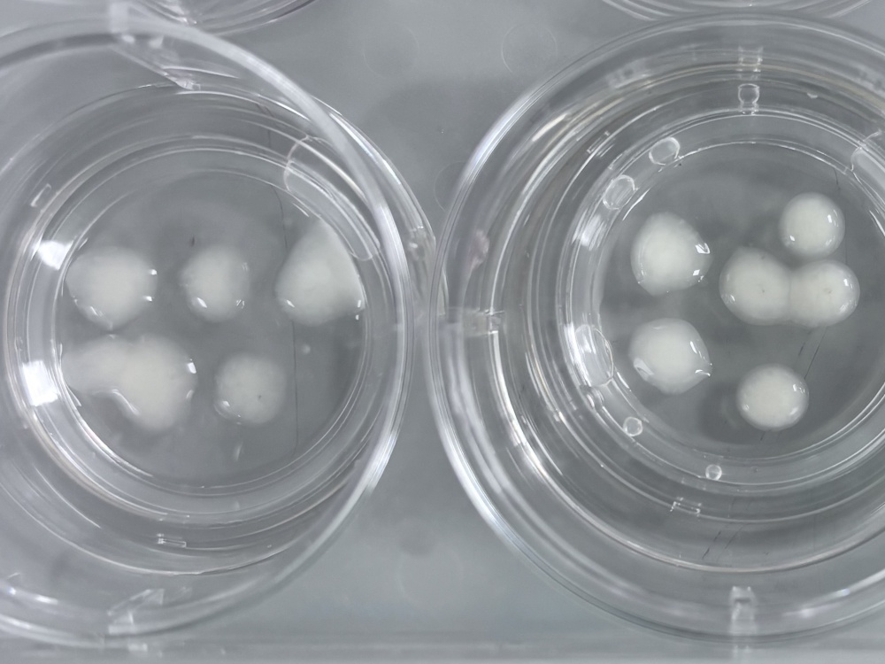
‘Mini brains’ might sound like something straight out of science fiction, but actually they’re becoming an important tool in medical research. Known as brain organoids, they’re small clusters of human brain cells grown in the lab.
They’re not real brains. They can’t think, feel or behave, but because they’re made from human cells, they can sometimes provide insights that are difficult to obtain from animal studies alone.
This new study, led by the University of Cambridge, used brain organoids to explore how one of the most common mitochondrial DNA mutations, called m.3243A>G, affects brain cells. This mutation is linked with MELAS, a mitochondrial syndrome that can involve seizures, stroke-like episodes, muscle problems, hearing loss and diabetes.
Why focus on the brain?
Mitochondria help produce the energy our cells need to work properly. The brain is one of the most energy-demanding organs in the body, so it can be particularly vulnerable when mitochondria aren’t working as they should.
People with mitochondrial disease can experience a wide range of neurological symptoms including seizures, stroke-like episodes, movement problems, developmental delay, learning difficulties and problems with balance or co-ordination.
Scientists still don’t fully understand why some brain cells seem to be more affected than others. This study set out to explore that question.
Why use organoids?
Studying the effects of mitochondrial disease in the human brain is difficult. Animal models have helped researchers understand some mitochondrial disorders, but there’s currently no animal model available for the m.3243A>G mutation.
That’s where organoids can help. Because they’re grown from human cells, they can carry the same disease-linked mutation found in people. They also allow scientists to study human brain cells in a three-dimensional, tissue-like environment.
How did researchers make the organoids?
The research team started with cells donated by people carrying the m.3243A>G mitochondrial DNA mutation.
In the laboratory, these cells can be turned back into stem cells. Stem cells are special because they can be encouraged to grow into many different types of cells, including brain cells.
Using this approach, the team grew brain organoids that carried different levels of the m.3243A>G mutation. This matters because mitochondrial DNA mutations often involve something called heteroplasmy.
Heteroplasmy means that a cell contains a mixture of healthy and altered mitochondrial DNA. The higher the proportion of altered mitochondrial DNA, the more likely it is that the cell may struggle to make enough energy.
By comparing organoids with low, medium and high levels of the mutation, the researchers could ask whether brain cells become more affected as the mutation level increases.
What did they find?
The team found that cells could cope with low and moderate mutation levels by adapting the way they produce energy. However, once mutation levels became too high, these protective mechanisms failed, leading to energy shortage and cellular stress. In simple terms, their mitochondria were less able to support normal energy production.
The cells appeared to shift towards alternative ways of making energy, suggesting they were trying to compensate for mitochondrial stress. But when the mutation level was high, some cells appeared unable to cope.
Importantly, not all brain cells were affected equally.
The most vulnerable cells were a particular type of neuron. Neurons are nerve cells that send and receive messages in the brain and nervous system.
The neurons most affected in this study are involved in long-distance communication. They send long connecting fibres to other parts of the brain and nervous system. Because they have so much ‘wiring’ to maintain, they need a lot of energy.
In organoids with high levels of the m.3243A>G mutation, these neurons showed signs of stress, damage to their long connecting fibres and increased cell death.
In additional to the organoid work, the same selective loss of neurons was also observed in postmortem brain tissue from a patient with MELAS, confirming that the organoids closely mimic human disease. One important next step will be to investigate whether this is a consistent finding in additional autopsy samples.
This could help explain why mitochondrial disease can cause serious neurological symptoms such as seizures and stroke-like episodes. If some of the brain’s communication cells are especially sensitive to mitochondrial dysfunction, their damage could have widespread effects.
What are the limitations?
Organoids are powerful tools, but they’re still simplified models.
A brain organoid is not a complete brain. It doesn’t have the full structure, blood supply, immune system, sensory input or long-range connections of a person’s brain. It also can’t show the full-body effects of mitochondrial disease, where many other organs and systems may be involved.
Organoids also tend to model aspects of early brain development rather than the full complexity of an adult brain. This matters because many people with mitochondrial disease develop symptoms over time, and age, environment and other genetic factors may influence how the condition manifests.
This means organoids are best thought of as one tool in the research toolbox. Their findings still need to be interpreted alongside other laboratory models, clinical studies and patient experience.
The bottom line
This study gives researchers a new way to investigate how the m.3243A>G mitochondrial DNA mutation affects human brain cells.
It shows that high levels of the mutation can disrupt mitochondrial energy production, and that some neurons may be especially vulnerable because of their high energy needs.
This research doesn’t change treatment for people affected by mitochondrial disease today. But what it does so is provide an important model for studying disease mechanisms in human cells, which is a vital step towards developing better therapies in the future.
You can read the full paper here.